Where is the xiphoid process of the sternum located photo
The xiphoid process, although small, is an important part of the human skeleton. Sometimes you may find that when you press on it, it becomes painful. Code for MBK-10 – M54.6 (pain in the thoracic spinal column). Negative feelings indicate diseases and malfunctions of the body.
xiphoid process
In the middle of the chest there is a small process that projects downwards. Because of its shape it is called xiphoid. In infants, it is a dense cartilage and is not connected to the sternum.
Until the age of thirty, the tissues of the appendix gradually harden, and after 30 years they begin to grow into the bones. The shape and size of the formation can vary greatly. The process sometimes has a small hole in the middle or has a forked top. It can be blunt or acute.
Location and functions of the xiphoid process
The xiphoid process (shown in the photo) is the lowest, smallest part of the chest. At first the formation is small, cartilaginous, triangular in shape. Then it gradually ossifies and fuses with the sternum. The process is located below its body and is attached using a fibrous connection. You can find the xiphoid formation by running your hand along the sternum - from the neck down to the end of the bones. The last one will be the shoot.
This is an important point of muscle attachment and is involved in the breathing process. The xiphoid formation connects the transverse and rectus abdominis muscles. During cardiopulmonary resuscitation, the appendix is used as a reference to determine the position for (indirect) cardiac massage. In this case, it is very important not to exceed the permissible pressure on the xiphoid formation, otherwise a puncture of the liver or sternum diaphragm may occur.
What does it mean if pain occurs when pressing on the appendix?
If the area of the xiphoid process of the sternum hurts when pressed, this may indicate the presence of a number of diseases or damage to nearby internal organs:
- stomach;
- hearts;
- lungs;
- gallbladder;
- pancreas.
In this case, pain occurs not only when pressed, but also with any slight exertion or overbite. Other reasons for the appearance of a negative symptom:
If pressure on the xiphoid process causes pain, it is important to pay attention to other associated negative signs. This is important for correct diagnosis.
Causes of xiphoid pain
The lower part of the sternum is covered by a fibrous plate. If it is not there, then the process may protrude somewhat. When pressure is applied to it, pain appears. Their causes can be various factors and diseases.
Pathologies and injuries
The pain may be caused by inflammation of the xiphoid process of the sternum. At the same time, when pressed, the sensations become stronger. Pain is also caused by a number of diseases:
- chondropathy;
- benign and cancerous neoplasms;
- osteochondrosis;
- hernias;
- diseases of any organs located near the xiphoid process;
- pathologies of the cardiovascular system.
Pain occurs when muscles are torn due to a strong blow. Injuries cause fractures and bruises. In this case, the pain can be very intense and intensify with breathing, sudden movements or coughing. After injury, negative feelings persist for a long time.
Hernia of the xiphoid process
When the xiphoid process thickens (bulges out), the pain may be caused by a hernia. It usually appears as a result of injury or is inherited. The xiphoid formation may have several openings, normally closed by a fibrous plate.
 If it is absent, a series of holes penetrates elements of nearby organs or fatty tissue. As a result, a pre-abdominal lipoma is formed. A true hernia of the appendix occurs rarely. It is characterized by bulging of the xiphoid formation and pain in the sternum. On palpation, the hard edges of the hernia and the contents of its sac are felt. The disease is treated with a simple surgical operation.
Tietze syndrome
Tietze syndrome can appear for no reason or as a result of:
- calcium deficiency;
- metabolic disorders;
- diseases of muscles and joints;
- hypovitaminosis;
- age-related changes;
- excessive loads;
- infections.
Tietze syndrome is an aseptic inflammation of cartilage tissue. It is characterized by pain at the junction of the chest bones and the upper ribs. Negative sensations can radiate to the appendix and occur when pressure is applied to it.
Slipping rib syndrome
Slipping rib syndrome is a condition in which pain occurs at the tips of the ribs or where they connect to the chest. Negative sensations intensify when pressure is applied to the xiphoid process. The disease appears due to recurrent subluxation of the cartilage. This leads to rib sliding (its excessive mobility). Then the bone touches the nerve, which causes pain.
Abdominal problems
They can appear as a result of diseases of the internal organs located in the peritoneum. In this case, the pain does not have a clear localization. It intensifies after pressing on the process. Diseases of the peritoneal organs include:
- pancreatitis;
- diseases of the esophagus;
- stomach ulcer;
- cholecystitis;
- gastritis.
Gastrointestinal pathologies can be distinguished from musculoskeletal pathologies in the presence of additional symptoms - nausea and vomiting, belching, heartburn. Sometimes constipation occurs. For diagnosis, a blood test is taken and an ultrasound is performed.
 Pain in the area of the xiphoid process can cause thoracic osteochondrosis, as well as hernias and protrusions of intervertebral discs. They gradually lose their elasticity. As a result, the tissues begin to bulge, compressing the nerve fibers, causing pain. It depends on the direction and size of the protrusion.
Other reasons
Other causes of pain in the xiphoid process include diseases of the respiratory organs - tuberculosis, pneumonia, bronchitis. Negative sensations sometimes arise due to benign and cancerous tumors, vascular pathologies, and fibromyalgia. The cause may be abnormal development (aplasia of the process) or xiphodynia (hypersensitivity of the xiphoid formation).
Pain may occur due to heart disease, in particular angina. During flatulence, gases create excess pressure. It also affects the xiphoid process, since the top of the colon is nearby.
Treatment
If pain occurs, you should consult a therapist. He will prescribe a series of tests and refer you for instrumental diagnostics, and, if necessary, for consultation with a gastroenterologist, traumatologist, surgeon or other specialists.
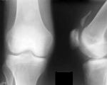
The xiphoid process is examined using:
- x-ray;
- FGDS.
Drugs are prescribed to eliminate symptoms (analgesics, antispasmodics, etc.). For musculoskeletal diseases, anti-inflammatory non-steroidal drugs, chondroprotectors, corticosteroids, and muscle relaxants are indicated. For severe pain, anesthetic blockades are placed. After eliminating the main symptoms, physiotherapy is prescribed.
If the cause of pain in the appendix is gastrointestinal pathology, then treatment is accompanied by a diet. Moreover, in case of chronic diseases, you need to adhere to it all your life. The treatment of cardiovascular pathologies includes cardiological and blood pressure-normalizing drugs. Some need to be taken for life.
 To eliminate pain in the area of the appendix, you can apply ice wrapped in soft cloth there. With xyphoidalgia, it is prohibited to do traction and straightening of the spine, exercise therapy.
Surgeries are performed only in cases where there is a threat to life or severe, prolonged pain. When treating a slipped rib, the diseased portion of the bone is removed. The reason for the operation is the presence of a large hernia.
Prevention
Prevention has a general and specific nature. In the first case, it is recommended:
- healthy eating;
- constant physical activity;
- exclusion of stress;
- maintain a sleep schedule;
- rejection of bad habits;
- compliance with the diet.
For specific prevention, doctors' instructions are followed. These include maintenance medications, avoiding heavy exercise, and following a diet. Additionally, physiotherapy is prescribed. Congenital abnormal structure of the chest requires constant monitoring.
Pain in the area of the xiphoid process can often indicate the presence of diseases. If you have this symptom, you should consult a doctor. Self-medication is unacceptable, since only a comprehensive diagnosis can identify the cause of pain.
Cure arthrosis without drugs? It's possible!
Get the free book “Step-by-step plan for restoring mobility of the knee and hip joints with arthrosis” and start recovering without expensive treatment and surgery!
Get the book
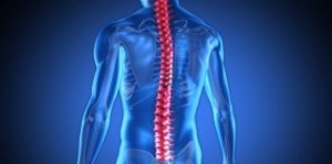
In the human body, despite its relative fragility, there are still effective structures that provide a protective function. All vital internal organs - the brain and spinal cord, heart, lungs - are hidden behind reliable bone formations. But if the skull or spinal canal have fairly stable dimensions, then the chest requires their constant change in the process of movement or breathing.
The anatomy of this formation is quite simple - its external supporting frame is formed by only three types of bones. But the volume is already determined by their total number - the sternum, twelve paired ribs and a similar number of vertebrae form the second largest cavity in the body. Also, the human chest is not only a supporting, but also a mobile formation, directly participating in the work of the lungs.
Mobility is given to it by a large number of joints - each rib and vertebrae has a separate connection to each other, as well as the strength of the surrounding muscles and ligaments. This combination of properties provides reliable protection for the heart, lungs and large vessels located inside the cavity formed. Therefore, damage to any part of the chest poses a threat to these vital organs.
Support structures
Before considering individual elements, you should pay attention to the general properties of this anatomical formation. Many people have difficulty imagining where exactly their chest is, pointing only to its upper part. Therefore, it is necessary to describe some of its external qualities:
- The upper border is located approximately at the level of the shoulder girdle, behind which the first pair of ribs is located. Since they are at the same level, a kind of bone ring is closed - an aperture.
- The lower part of the formation does not form a smooth border - it runs in an oblique direction. In the lateral and posterior sections, the chest reaches the level of the lower back, and in the abdominal area, the line rises along the lower edge of the ribs.
- Normally, the supporting structures are formed in the form of a slightly compressed and truncated cone, with the base directed downward. This structure is due to the shoulder girdle at the top, which requires some space for mobility.
The formation has elasticity due not only to ligaments and muscles, but also to the type of bones included in its composition - the ribs, sternum and vertebrae are formed mainly by spongy tissue.
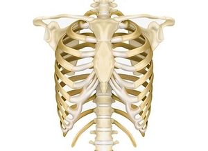
Sternum
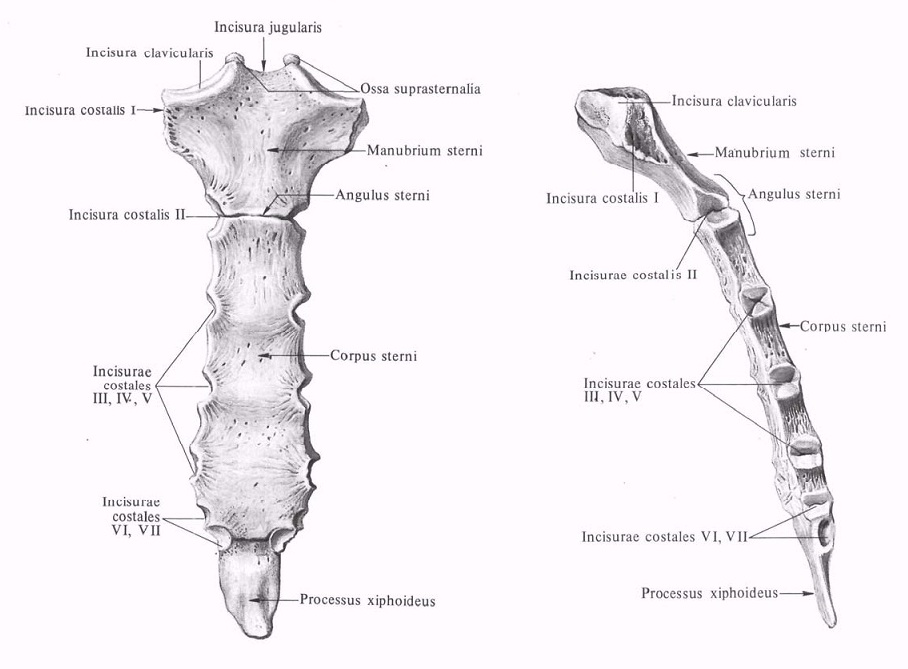
This structure forms the anterior rib cage and is the attachment site for most costal cartilages. Externally, it is a wide and slightly concave plate, consisting of three sections. Together they are connected by dense strands of connective tissue that form sutures. This structure is due to the need for slight stretching that occurs during movement and breathing.
The anatomy of this bone is considered from the point of view of each department, which has its own characteristics. But together they still form a strong and indivisible structure:
- The uppermost and widest part is the handle - in shape it resembles an inverted trapezoid, attached from below to the body of the sternum using a suture. On top it has paired symmetrical notches in which the sternal ends of the clavicles are located. In the same area, bundles of the largest muscle of the neck, the sternocleidomastoid, depart from it.
- The middle section is the body - it usually connects to the handle not directly, but at a slight angle. This feature is due to the fact that the chest narrows slightly in the upper segment. This section of the bone is the longest, representing an elongated rectangle.
- The lower part of the sternum is considered to be the xiphoid process, a small movable bony segment. Its structure is very variable - each person has its own size and shape. It can be felt just below the body of the sternum in the area of the junction of both costal arches.
This bone structure not only performs supporting functions, but is also one of the important hematopoietic organs in an adult.
Ribs
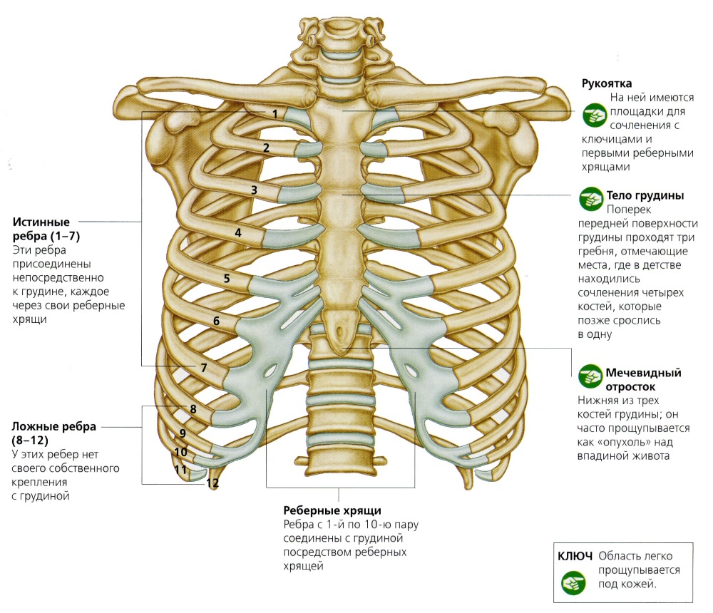
The anatomy of the ribs is quite simple - it is a thin bone curved laterally. At its posterior end there is a rounded surface necessary for attachment to the spine. In front, the rib, on the contrary, ends with a sharp edge, from which a cartilaginous outgrowth extends to the sternum.
It is difficult to find such a large number of identical bones in the human musculoskeletal system. Even the vertebrae in different sections have characteristic features that make it possible to separate them from their “brothers”. And almost all ribs differ in appearance only in size, since their anatomy obeys its own rules. Therefore, it is necessary to consider individual groups and elements that stand out from the crowd:
- Only those ribs that are attached directly to the sternum with their cartilage are considered true ribs. Usually these are the top seven pairs - they have a relatively straight direction.
- Next comes a group of false ribs - usually about two or three on each side. Their cartilages are no longer fixed to the sternum, but to the surface of the overlying similar bone.
- The eleventh and twelfth pairs are considered free - they are held in a transverse position only by the surrounding soft tissues. Their anterior edge is located in the area of the lateral borders of the abdomen.
The ribs are given simultaneous strength and elasticity by their special structure - their upper and outer edges are formed by thin compact bone, and their inner and lower sections are formed by spongy substance.
Spine
![]()
In addition to the bones listed, the chest also has a main supporting element - the thoracic segment of the spinal column. Thanks to the special structure of the joints between the ribs and the spine, they work together during breathing and movement:
- The main joint is the costovertebral joint - it is located in the depression, which is located between adjacent vertebrae. The head of the rib is securely attached to it with the help of ligaments. Due to the anatomy of the surrounding tissues, movements in these joints are always joint.
- For additional support, a costotransverse joint is formed a little further, which does not play a big role in the mobility of the chest. Its purpose is to prevent excessive displacement of the ribs up and down. It is formed between the costal tubercle and the inner surface of the transverse process of the vertebra.
With any rotation of the torso or tilt, the chest stretches along with the spine, providing a person with freedom of movement.
Soft fabrics
![]()
In addition to the external bone frame, which plays a predominantly supporting role, there are also dynamic elements. The structure of the human chest also includes a large number of muscles involved in the act of breathing. Based on localization, they can be divided into the following groups:
- The most important anatomical structure separating the chest cavity from the abdomen is the diaphragm. It is a wide and flat muscle, shaped like a dome. When it contracts and relaxes, a significant change in pressure inside the chest cavity occurs, which ensures proper functioning of the lungs.
- Also, intercostal muscles - narrow muscle cords that connect the lower and upper edges of adjacent bones - are actively involved in breathing. In humans, they consist of two differently directed layers - the contraction of each of them provides inhalation or exhalation.
- Some muscles of the shoulder girdle are attached to the surface of the ribs, providing their mobility. These include the pectoralis major and minor, subclavian and serratus anterior muscles. With calm breathing, they practically do not work, but with heavy loads, their contraction allows you to more effectively expand the chest.
The abdominal muscles can also be attributed to the respiratory muscles - they change intra-abdominal pressure, indirectly affecting the functioning of the lungs.
Thoracic cavity
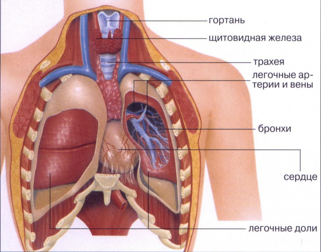
Inside, the resulting space is densely filled with internal organs covered with special shells. Based on this feature, it can be divided into the following parts:
- On both sides are the lungs, covered with sheets of pleura - a tissue that ensures their free movement. It consists of two sheets, between which there is a little liquid that prevents them from rubbing against each other.
- The anterior mediastinum is located just behind the sternum - in an adult there are only lymph nodes, blood vessels and adipose tissue. And in children there is an important organ of immunity - the thymus gland.
- The middle mediastinum is formed by the pericardial cavity - it houses the heart, and large vessels extending from it. It also contains the terminal section of the trachea and the main bronchi leading to the lungs.
- The posterior mediastinum is completely filled with anatomical formations - the esophagus, the lymphatic duct, as well as large nerve trunks and veins pass between the heart bag and the spine.
It is these important formations that are protected by a strong and elastic frame of the chest, ensuring their smooth operation. Without the protection and support of bones and muscles, they would easily be subjected to life-threatening injuries.