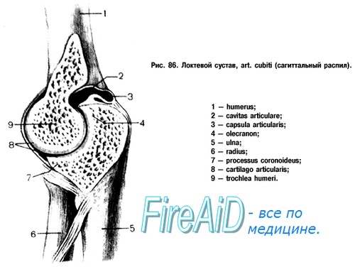
elbow joint
Elbow joint, articulatio cubiti. Three bones articulate at the elbow joint: the distal end of the humerus and the proximal ends of the ulna and radius. Articulating bones form three joints enclosed in one capsule (complex joint): humeroulnar, art. humeroulnaris, brachioradialis, art. humeroradialis, and proximal radioulnar, art. radioulnaris proximalis. The latter functions together with the distal articulation of the same name, forming a combined joint.
Shoulder joint is a block-shaped joint with a helical structure of the articular surfaces. The articular surface from the side of the shoulder is formed by a block, trochlea; the recess located on it (guide groove) is not located perpendicular to the axis of the block, but at a certain angle to it, as a result of which a helical stroke is obtained. Connected to the block incisura trochlearis ulna, which has a comb corresponding to the same notch on the block of the humerus.
Shoulder joint formed by articulation capitulum humeri with a fossa on the head of the radius and is spherical in shape, but in fact, movement in it occurs only around two axes, possible for the elbow joint, since it is only part of the latter and is connected with the ulna, which limits its movement.
Proximal radioulnar joint consists of interconnected circumferentia articularis radii and incisura radialis ulnae and has a cylindrical shape (rotary joint of the first type). The articular capsule on the humerus covers behind two-thirds of the cubital fossa, in front of the coronary and radial, leaving free epicondyles. On the ulna it is attached along the edge of the incisura trochlearis. It is fixed on the beam along the neck, forming a protrusion of the synovial membrane in front - recessus sacciformis. The capsule is free in front and behind, but there are auxiliary ligaments on the sides: lig. collateral ulnare from the ulnae and lig. collateral radiation from the side of the beam, located at the ends of the frontal axis and perpendicular to it. Lig. collateral ulnare starts from the medial epicondyle of the humerus and is attached along the entire medial edge of the incisura trochlearis ulnae. Lig. collateral radiale starts from the lateral epicondyle of the shoulder, covers the head of the radius in front and behind with two legs and is attached at the anterior and posterior edge of the incisurae radialis ulnae. The gap between both legs is occupied by fibrous fibers that curve around the neck and head of the ray in an arcuate manner, without fusing with them. These fibers are called lig. annulare radii. Due to this position of the annular ligament, in a horizontal plane, perpendicular to the vertical axis of rotation, the ligament directs the movement of the beam around this axis and keeps it without interference for rotation.
Movements in the elbow joint are of two kinds. First, it performs flexion and extension of the forearm around the frontal axis; these movements occur at the articulation of the ulna with the block of the humerus, and the radius also moves, sliding along the capitulum. The amount of movement around the frontal axis is 140°.
The second movement consists in the rotation of the radius around the vertical axis and occurs in the glenohumeral joint, as well as in the proximal and distal radioulnar joints, which, therefore, represent one combined rotational joint. Since the brush is connected with the lower end of the beam, the latter follows the radius when moving. The movement in which the rotating radius crosses the ulna at an angle, and the hand turns with the back side forward (with the arm lowered), is called pronation, pronatio. The opposite movement, in which both bones of the forearm are parallel to each other, and the hand is turned with the palm forward, is called supination, supinatio. The range of motion during pronation and supination of the forearm is approximately 140°.
The ability of the bones of the forearm to pronate and supinate, which was in its infancy in animals, improved in primates in connection with climbing trees and an increase in grasping function, but reached its highest development only in humans under the influence of labor.
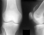
On radiographs of the elbow joint region, a simultaneous image of the distal humerus and the proximal forearm bones is obtained. On the back and side images, all the details of these departments described above are visible. In the side view, trochlea and capitulum humeri are layered on top of each other, as a result of which the shadows of these formations look like concentric circles. The “X-ray joint spaces” of articulatio humeroulnaris, articulatio humeroradialis, art. radioulnaris proximalis.
On the posterior radiograph, the gap of the glenohumeral joint is especially clearly visible; on the lateral picture, the gap of the glenohumeral joint is traced throughout.
The elbow joint receives arterial blood from rete articulare formed aa. collaterals ulnares superior et inferior (from a. brachialis), a. collateralis media and collateralis radialis (a. profunda brachii), a. recurrens radialis (from a. radialis), a. recurrens interossea (from a. interossea posterior), a. recurrens ulnaris anterior et posterior (from a. ulnaris). Venous outflow through the veins of the same name occurs in the deep veins of the upper limb - vv. radiales, ulnares, brachiales. The outflow of lymph occurs through the deep lymphatic vessels in the nodi lymphatici cubitales. The innervation of the joint capsule is provided by n. medianus, n. radialis, n. ulnaris.