Human scapula. Anatomy of the human scapula
The system of support and movement, which includes bones, muscles and ligaments, functions in the human body as a whole. The skeleton, formed by a special type of connective tissue cells - osteocytes, consists of several sections. It includes the skull, spine, chest, free limbs and belts that connect the bones of the upper and lower limbs with the spine.
In this work, we will focus on the structure of the human scapula, which, together with the clavicle, forms the girdle of the upper limbs. We will also determine its role in the skeleton and get acquainted with the most common developmental pathologies.
Features of the structure of flat bones
The supporting apparatus contains several types of mixed and flat. They differ from each other both in appearance and internal anatomical structure. For example, a compact bone substance may have the form of two thin plates, between which, like a layer in a cake, there is a spongy tissue penetrated by capillaries and containing red bone marrow.
It is this structure that the sternum, cranial vault, ribs, pelvic bones and human scapula have. It best contributes to the protection of the underlying organs: lungs, heart and large blood vessels from mechanical shock and damage. In addition, a large number of muscles that perform static and dynamic work are attached to the vast flat surface of the bone with ligaments and tendons. And the red bone marrow, located inside a flat bone, serves as the main hematopoietic organ that supplies the formed elements: erythrocytes, leukocytes and platelets.
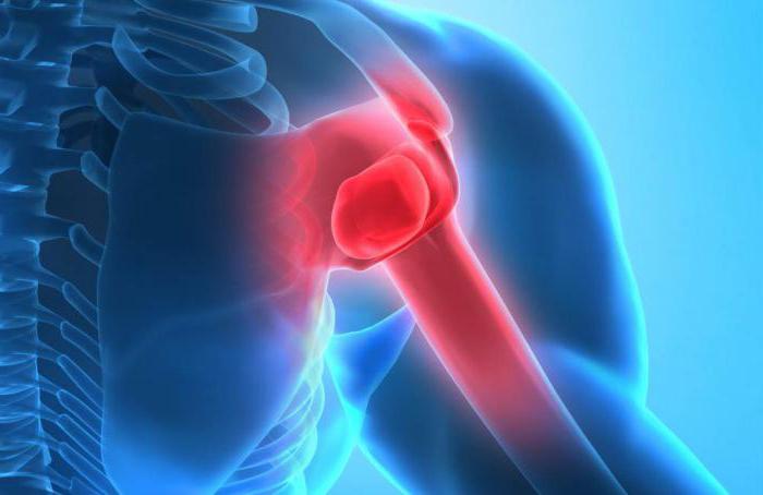
Anatomy of the human scapula
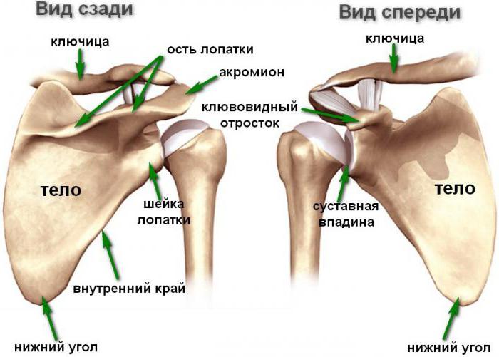
The bone has the shape of a triangle touching the posterior surface of the sternum. Its upper part has a cut edge, the medial section is turned towards the spine, the lateral angle contains the articular cavity. It includes the head of the tubular humerus. Another element of the upper limb belt is the clavicle, which is connected to the scapula with the help of the acromioclavicular joint. The axis passing along the posterior surface of the scapula reaches the lateral surface, passing into the acromion. It has a junction with the clavicle in the form of an articular surface. A more complete picture of the anatomical features of flat bones is given by the photo of the human scapula, presented below.
In embryogenesis, bone is formed from the mesoderm. In a newborn, the ossification of the scapula is not complete and osteocytes are found only in the body and spine, the rest has a cartilaginous structure (endochondral type of ossification). In the first year of a child's life, points of ossification appear in the coracoid process, later in the acromion - the lateral end of the scapula. Complete ossification is completed by the age of 18.
How muscles attach to the shoulder blade
The main way to connect bones and muscles in the musculoskeletal system is with the help of tendons.
Thanks to collagen fibers, which are the end part of the biceps, the biceps brachii is attached to the tubercle located above the upper edge of the glenoid cavity of the scapula with its long head. The lower edge has the same bumpy surface, to which, with the help of a tendon, a muscle is attached that extends the arm in the shoulder joint - triceps
Thus, the human scapula is directly involved in the flexion and extension of the upper limb and the maintenance of the muscular corset of the back. The bones of the girdle of the upper limbs - the collarbone and shoulder blades have a common system of ligaments, however, the scapula has three own ligaments that are not related to the shoulder and acromioclavicular joints.
The meaning of the coracoid process
A part of the bone extends from the upper edge of the scapula, which is the remnant of the coracoid of vertebrates and is called the coracoid process. It is located above the shoulder joint like a visor. The short head of the biceps, as well as the beak-shoulder and pectoralis minor muscles, are attached to the process with the help of tendons.
Being part of the scapula - a human bone that directly forms the girdle of the upper limbs, the coracoid process is involved in the work of the antagonist muscles: the biceps and triceps, and its connection with the muscles of the shoulder ensures the abduction of the upper limb to the sides and up. As you can see, the coracoid process is of no small importance in the structure of the scapula. What is its anatomical origin?
Coracoid and its role in the phylogeny of vertebrates
Earlier, we focused on the fact that the paired clavicle and scapula enter the upper limb girdle. Man is distinguished from other vertebrates, for example, from birds, reptiles, fish or amphibians, by the reduction of the crow bone - the coracoid. It is associated with the release of the upper limb from physically complex and diverse motor functions in the form of running, flying, swimming or crawling. Therefore, the presence of a third bone in the girdle of the forelimbs became impractical. The crow bone in humans was reduced, only a part of it was preserved - the coracoid process, which became part of the scapula.
Pathology of the bones of the girdle of the upper limbs
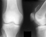
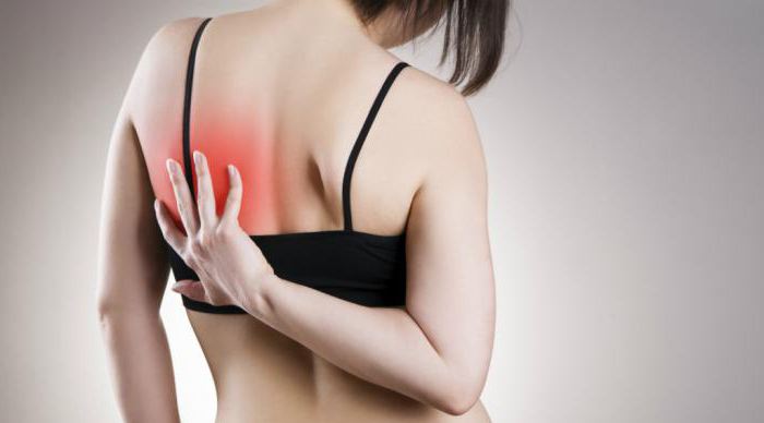
The most common anomalies in the structure of the human scapula arose as a result of both violations of organogenesis during fetal development, and in the form of complications after dystrophic muscle damage or neuroinfections. These include, for example, the syndrome of the pterygoid scapula, which is determined both during an external examination of the patient and on an x-ray.
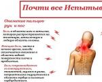
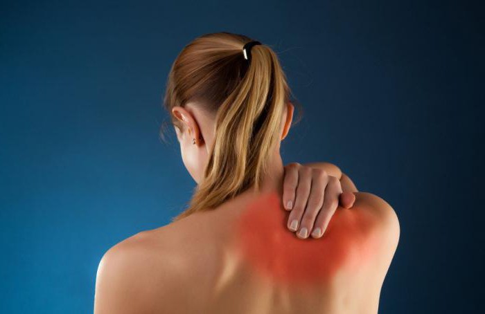
The disease is accompanied by debilitating pain in the shoulder and behind the sternum as a result of a rapidly developing neuropathy. Remission occurs when therapeutic and preventive measures are observed: dosed physical activity, massage, special exercises for the muscles of the shoulder and back.
Another pathology is congenital high standing of the scapula (Sprengel's disease). This anomaly is combined with a violation of the structure of the vertebrae, anatomical defects of the ribs, for example, their fusion or partial absence. There are two forms of the disease: unilateral and bilateral violation of the symmetry of the shoulder blades.
So, with a bilateral lesion, the left shoulder blade is located higher than the right one. The anomaly is dangerous by the degeneration of myocytes into the main and rhomboid - large and small. A positive prognosis can be expected from a surgical intervention performed on a child under 8 years old; at a later age, surgery is not resorted to due to the high risk of complications, limited to therapeutic exercises and massage.