Human foot structure: schemes and diseases of organs and bones, muscle points with Photos and treatment
Human evolution has made the foot a unique and complex mechanism that performs springing and balancing functions, providing shock mitigation during movement.
Thanks to the limbs, a person got the opportunity to move, keep balance, and resist movement.
There are 26 bones in the foot and all of them are connected into one mechanism by ligaments and joints.
In addition, there is a huge amount of muscle tissue and tendons.
Bones
The foot and hands are similar in structure. Anatomy divides the foot into the following bone sections:
tarsal
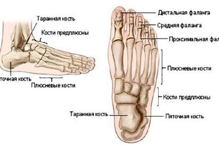 Includes 7 bones. The most bulky are ram and heel. The talus is located between the lower leg and refers more to the ankle. This includes:
- - club-shaped;
- - scaphoid;
- - sphenoid bone.
Metatarsus
This is a collection of five bones resembling tubules in shape. This department is average and is responsible for the functioning of the fingers and the correct location of the arch. Bones ending in joints lead to the beginning of the fingers.
Distal
It has 14 bones. Each finger has 3 bones, except for the thumb, which has only two. Between the bone formations are joints for mobility.
Thanks to this zone of the foot, the human body keeps balance and can move. Interestingly, in case of loss of hands, the toes perform a replacement function.
The joints are located between the bones. In addition, the foot contains muscles, ligaments, nerves, and blood vessels.
How are the bones located?
Bones require more detailed consideration, since they are the main component of the foot.
The heel bone is the most powerful
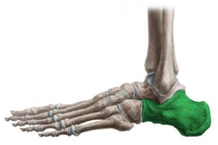 It is located in the back and carries a huge load. Despite the fact that this part has nothing to do with the ankle, it plays a large role in the distribution of pressure. The shape of the calcaneus resembles a triangle in three dimensions with a long axis.
The role of the connector between the calcaneus and the talus is performed by the joints. A strong connection of these two bones is necessary to give the foot a normal shape. The back of the bone holds the Achilles tendon. This place can be found on a small ledge. And the lower part is a support when walking on the surface of the earth.
On the front part, you can find a tubercle where the scaphoid bone and the joint are connected. On the surface, you can see many protrusions and vice versa - depressions. These are the places where blood vessels, muscles, nerves, ligaments are attached.
The talus is several times smaller than the calcaneus
But massive and is part of the ankle. She is turned to the heel. It mainly consists of cartilage and, surprisingly, it does not hold anything other than ligaments. Its surfaces, consisting of 5 pieces, are lined with a thin layer of hyaline cartilage.
This bone is made up of the following parts:

Despite the power of the bone, it is often injured or ill.
cuboid
You can find it on the outside of the foot at the outer edge. Located behind the 4th and 5th metatarsals. The shape is a cube, hence its name. Behind comes into contact with the calcaneus, and that is why it has a saddle shape and calcaneal process.
Scaphoid
 It is located directly on the foot at the inner edge.
Its ends are flattened, the upper part can bend, and the lower part is sunken.
Thanks to the joints, it interacts with the talus and serves as a foot shaper.
wedge-shaped
Consists of three bones:
- - medial, it is the largest;
- - intermediate, smallest;
- - lateral - middle.
They are all small and located quite close to each other. They have the metatarsal bones in front and the navicular bones behind. The entire system is strong and rigid, forming a solid foundation for the foot.
metatarsal bones
They are curved tubes. They have the same structure and carry out similar functions in both young and adult years. The bends of the bones give the arch the desired position. If you look at the surface, then it is characterized by tuberosity, due to the connection of ligaments, joints and muscles.
phalanges
The same as on the fingers. The difference is only in size. The thumb is assembled from 2 phalanges, and the shape is much thicker due to the resulting load when walking. The rest consist of three phalanges and are much thinner and shorter.
joints
What are joints made of?
The feet are distinguished by the presence of a large number of joints that play a reducing role between the bones. If we compare them in size, then the largest is the ankle joint, which connects three large bones together. This allows a person to raise and lower the foot, to make rotational movements. The remaining joints are much smaller, but in fact their function is similar. They give you the flexibility you need.
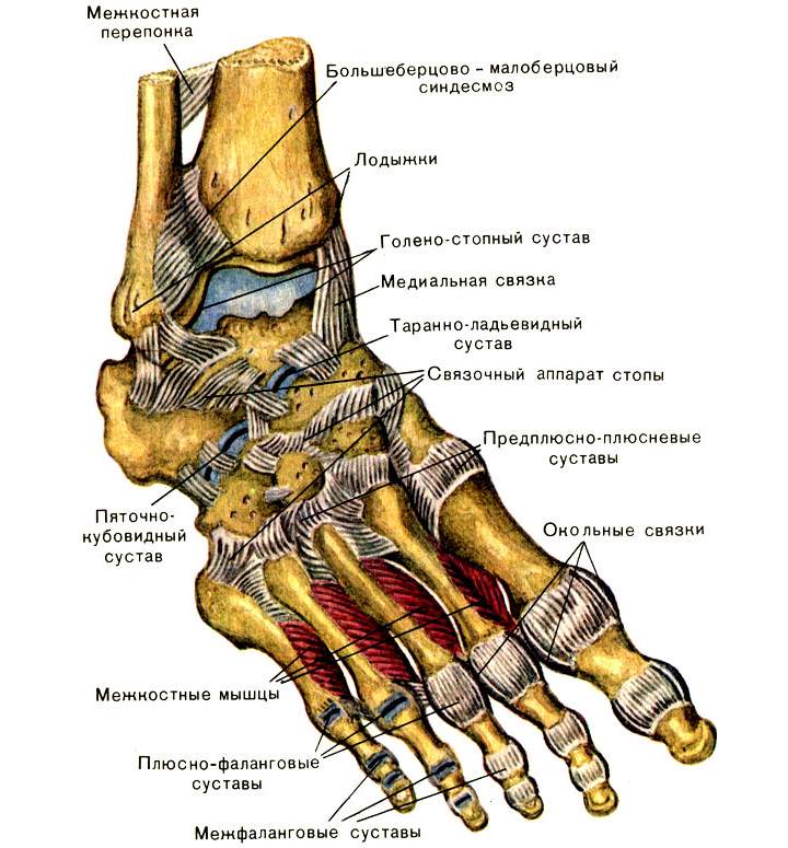 Let's say a little about the ankle joint. It includes a large talus and two smaller tibias that include the ankles. The edges of the joint are attached with strong ligaments, and it is securely connected to the cartilage.
A huge role is played by the transverse or subtalar joint. It is inactive, but connects as many as three bones - the scaphoid, talus and calcaneus. For more reliable fixation, participation in the connection of ligaments is provided.
The subtalar joint is helped to form the arch by the cuboid and heel joints. Sometimes such a connection is called the Greek cavity, and in medicine it was called the talonavicular joint.
One of the most important joints is the metatarsophalangeal. They take part in every movement of the human body.
The least significant are the joints on the navicular and sphenoid bones.
Bundles
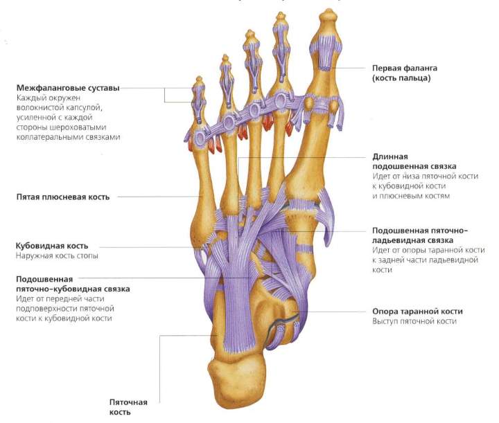 In first place in importance is the plantar ligament. It originates from the calcaneus and ends at the origin of the metatarsal bones.
The ligament is distinguished by a large number of branches that carry the fixing function of the longitudinal and transverse arches.
Such a connection is responsible for the correct condition of the arch throughout a person’s life.
Smaller ligaments are needed to strengthen the skeletal system and joints. Thanks to them, the human body is able to maintain balance and load during movements.
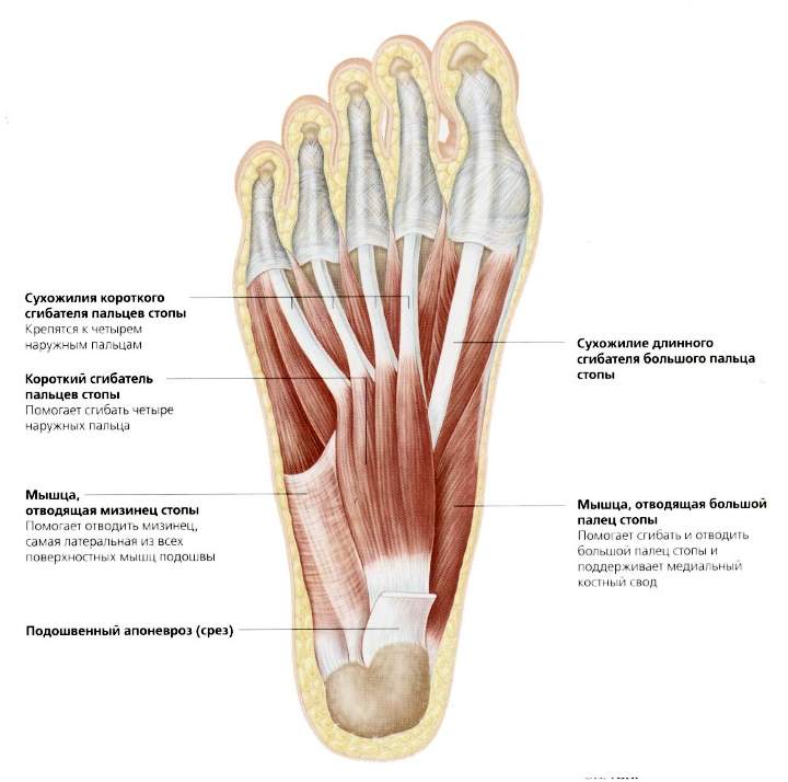
muscles
The foot can move only with the help of muscles. They are everywhere - in the area of the foot, lower leg and ankle. The muscular structure of the lower leg provides movement of the feet during walking and in an upright position.
 The anterior part consists of the extensor longus muscle group and the tibialis muscle. Thanks to them, the phalanges on the legs can be bent and unbent.
The long and short fibulas provide lateral flexion of the foot and pronation.
A very bulky muscle group is located in the back. These muscles are made up of several layers. This includes the following muscles:
- triceps, including gastrocnemius and soleus;
- finger flexor;
- plantar;
- tibial (partial).
The sole during the work of this muscle group is bent with the help of the Achilles tendon. And muscle tissue helps with bending and unbending the fingers.
For the movement of four fingers, not taking into account the big one, the extensor of the short type, which belongs to the dorsal muscle group, is responsible. Small muscles on the foot allow it to perform the functions of abduction, flexion.
Vascular and nervous systems of the foot
Blood
In order for blood to flow to the feet, tibial arteries are provided in front and behind. They stretch along the very foot on the sole. Small connections and circles depart from these large arteries.
When the foot is damaged, one of the circles is disrupted, but the others continue to provide the necessary blood flow to the limbs.
The veins on the back side are responsible for the outflow. They look intertwined and provide blood to the great and small saphenous veins in the lower leg.
Nerves
They form an integral part of the normal functioning of the human foot. They are responsible for sensations:
- - pain;
- - vibrations;
- - touch;
- - cold or warm.
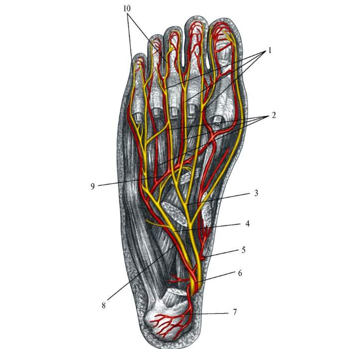 Nerve signals, leaving the CNS along the gastrocnemius, peroneal, superficial and tibial nerves, reach the spinal cord and are processed there.
Nerves transmit a signal to the muscles, being essentially reflexes - voluntary or involuntary (independent of human will). Involuntary include the work of the glands (sebaceous and sweat), vascular tone.
As for the skin, there are several zones on the foot that differ in density, structure, and elasticity. For example, the leather of the sole is high density, while the heels are thick. Initially, the skin of the palms and feet are the same, but over time and with increasing loads, additional layers appear. The back of the foot is smooth and elastic, with nerve endings.
Drawing a conclusion, we can say that nature has done everything so that the foot can withstand tremendous pressure.
Foot diseases
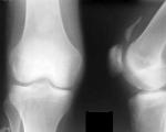
The foot is regularly subjected to loads, either static or impact. Injuries are common for her. Almost always accompanied by pain, an increase in some of the epiphyses, swelling, curvature. You can identify the pathology on an x-ray.
Arthrosis
This is a disease in which cartilage loses its elasticity. Often this disrupts metabolic processes. There is pain, crunch, swelling.
Causes of arthrosis:
- - infectious diseases;
- - allergies;
- - systemic diseases - lupus erythematosus, scleroderma;
- - tuberculosis;
- - syphilis;
- - dislocation or injury.
Often you can find arthrosis of the first toe.
The disease develops in 3 stages:
- At first, pain occurs, but disappears after rest. Sometimes the deviation of the thumb becomes noticeable. There is a crunch when moving.
- To dull the pain, painkillers and anti-inflammatory drugs are taken. The finger is already bent strongly and it becomes impossible to pick up shoes.
- The pain does not go away even when taking analgesics. The deformity extends to the foot, there is a problem with walking.
Arthrosis also strongly loves the ankle, deforming the joint and affecting the cartilage.
This disease is treated conservatively only at an early stage. Then you will need surgical intervention - arthroplasty, resection, arthroplasty.
flat feet
There are congenital or acquired flat feet. Reasons for the appearance:
- - excess weight;
- - heavy loads;
- - diseases of the nerve endings;
- - injuries;
- - Wrong shoes
- - transferred rickets or osteoporosis.
Flat feet come in two forms:
- Transverse - with a decrease in the height of the arch, when the heads of the metatarsal bones are in contact with the ground.
- Longitudinal - that is, the entire foot has contact with the ground. Increased fatigue in the legs, pain.
Arthritis
A joint disease that affects the entire human body. There are primary and secondary arthritis. The causes of the appearance are the same as with arthrosis. Symptoms include:
- - pain;
- - deformity of the leg;
- - swelling, redness;
- - fever, rash, fatigue.
Treatment methods depend on the underlying cause of the disease and can be physiotherapy, medication, manual, etc.
Clubfoot
As a rule, it occurs from birth. The reason is a subluxation of the ankle joint. Acquired clubfoot is the result of trauma to the lower extremities, paralysis, paresis.
Disease prevention
Preventing the development of diseases is much easier than treating. Prevention includes:
- performing special strengthening exercises;
- sparing sports - cycling, skiing, swimming;
- wearing comfortable shoes made from natural materials;
- walking on pebbles, sand, grass;
- the use of special orthopedic insoles;
- resting the legs.