The structure, functions and features of the shoulder joint
The shoulder joint is one of the largest joints in the human musculoskeletal system. The joint is formed by a specific mechanism: the head of the shoulder in the form of a ball surrounded by ligaments and muscles. All this gives a strong strength, but also a great vulnerability of the structure. The shoulder joint during human life is subject to significant physical stress.
The shape of the joint makes it possible to perform not only vital movements for the human body, but also to achieve high achievements in sports and work. The shoulder must function correctly. And for this it is necessary to lead a healthy lifestyle, properly rest, eat well and contact a specialist in a timely manner in case of pain or damage.
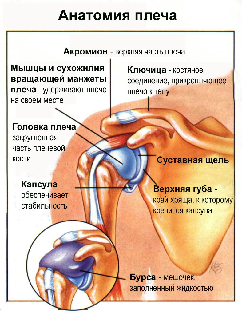
Shoulder Anatomy
Each joint of the human skeleton is formed by the articulation of two or more bones with the help of cartilaginous, connective tissues, ligamentous apparatus and muscles. The shoulder joint, in fact, is formed by a spherical joint that includes the scapula and humerus in its structure. Above the joint is an elastic capsule. The shoulder is strengthened by ligaments and muscles.
The anatomical features of the articulation provide an opportunity for interacting surfaces to move away from each other and return to their original position without damaging the integrity of the joint capsule.
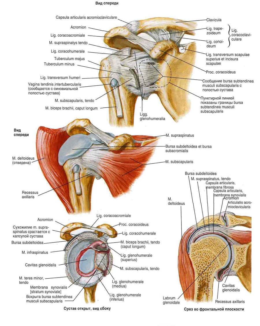
The shoulder joint is formed by the following parts of the bone skeleton: the head of the humerus and the scapular cavity. The shape of the ball is present at the bone of the shoulder, and at the cavity the shape is even in the form of a saucer. Such forms and the presence of hyaline cartilage make the combination of the bones of the shoulder girdle together with the scapula mobile. Cartilage has the form of a gel, which is formed by minerals and substances of organic origin, but it contains 80% water. The articular lip helps to balance the different surface dimensions. This element of the joint is formed by fibrocartilaginous tissue, which contributes to the excellent interaction between the scapular cavity and the shoulder.
The capsule is fixed at the end of the cartilaginous lip and scapular cavity. On the other hand, on the humerus, the capsule is well fixed along the anatomical neck. From below, it has a thin structure, but above a more thickened structure due to the tendons of different types of muscles that are woven into the capsule.
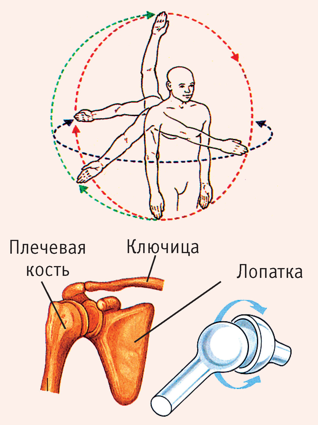
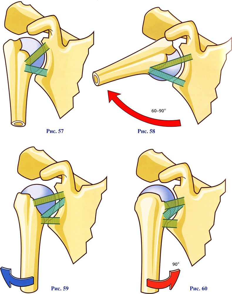
The main function of the shoulder girdle is to balance the movement of the arms during the increase in scope. That is, the mechanical ability of the shoulder girdle makes it possible to move the limbs in different projections at a large angle. At the same time, a strong attachment of the humerus (freely moving) and scapula (conditionally mobile) is given.
Hondrocream is the perfect solution for joint problemsThe structure of the shoulder joint makes it possible to carry out various movements with the upper limbs in a wide range: rotational, flexion, abduction, extension and adduction actions.
Motor ability of the shoulder joint
Movements with the shoulder girdle involved lead to the fact that the muscles gradually begin to displace the capsule. This is what prevents it from being infringed among the bone joints. The capsule is a bridge passing through the groove where the tendon fibers of the head of the muscle (biceps) are located. The fibers of this muscle originate from the end of the lip of the joint and from above the tubercle, and then stretches to the intertubercular groove. The muscle passes through the shoulder, where it is covered by a synovial membrane. The latter continues upward from the tendon fibers and passes into the capsular synovial membrane.
Features of the motor dynamics of the joint
Three ligaments are located on top of the capsule, attached in the region of the anatomical neck of the shoulder bone and cartilaginous lip. Ligaments help to strengthen the cavity of the capsule in front. Another shoulder contains a strong coraco-brachial ligament. It is similar to the fibrous tissue of the capsular layer, which is located from the large tubercle of the shoulder to the coracoid process.
The coracoacromial ligament is located over the articular joint of the shoulder. The arch of the shoulder is formed by this ligament, the coracoid and acromial processes. The arch contributes to the protection of the joint from above, makes a gradual retraction of the shoulder, lifting the limb forward and to the sides above the belt. At the moment when the arm rises above the waist, the work of the shoulder blades begins.
The structure of the bone in the shoulder
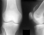
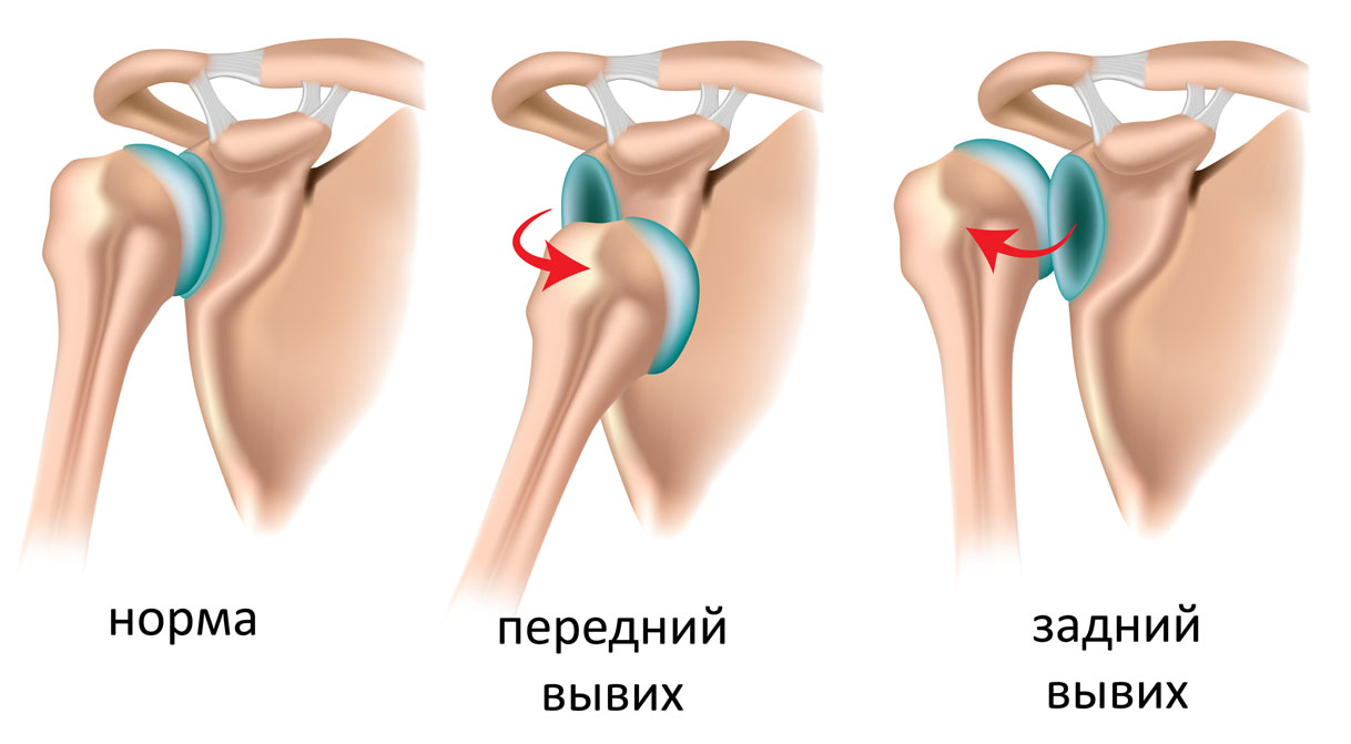
The main movements in the joint of the shoulder are performed using the head, located deep in the scapula. The shoulder joint is under heavy load. Because of this, inflammation and structural wear of the bone is quite common. To establish the diagnosis, the doctor may send for an X-ray examination. The resulting photo will allow you to accurately assess the condition of the joint.
Often there are such diseases of the articular joints as: congenital, traumatic, inflammatory and degenerative. Traumatic injuries include fractures, dislocations and subluxations. Degenerative damage includes arthrosis of the joint, during which thinning of the cartilage and bone tissue occurs, and there is a loss of the ability to move. Arthrosis occurs in older people. This may be due to metabolic disorders, frequent traumatic injuries, and a decrease in the intensity of blood supply to the osteoarticular system. Congenital pathologies are joint dysplasia (lack of full development of bone structures). Inflammatory diseases include arthritis obtained after an injury or as a result of systemic processes of an infectious type. Such violations must be treated, as they are dangerous for the development of serious complications.
Ligament mechanism of the shoulder
The most important element of the ligamentous mechanism is formed by the rotator cuff. This formation includes the following muscles of the shoulder joint: round small, infraspinatus, subscapular and supraspinatus. These muscles prevent injury and displacement of the head of the bone during the mobility of large muscles, namely: dorsal, biceps, deltoid and pectoral.
Shoulder ligaments do not have the ability to stretch strongly during heavy loads. This is what causes them to break. If a person does not exercise and moves little, then his muscles and shoulder joint will be fragile elements. This is due to the fact that such people have reduced blood supply, insufficient supply of nutrients to the joint, which leads to frequent injuries.
You should also not be zealous with excessive physical exertion, as this will lead to fatigue. The following tendon diseases may also appear and muscles may be injured:
- Ligamentous sprain after any injury contributes to a large loss of motor capabilities of a person with his hands. If treatment is not carried out, an inflammatory process will develop, which can spread to the tissues around.
- Periarthritis of the joint, that is, the process of inflammation in the tendons. This human disease is common, and it occurs after an injury: a bruise or a fall, or after heavy loads.
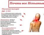
All injuries and pathologies of the shoulder joint include pain, which can be of varying degrees. Painful sensations are of very strong intensity and stop the motor abilities of the hand. All this is a protective mechanism, which is provided by the functions of the radial, thoracic, axillary and subscapular nerves, which ensure the conduction of signals through the joint. The pain syndrome leads to restriction of movements in the damaged articular joint, which provides an opportunity for inflamed and damaged tissues to recover.
It is worth paying attention to the fact that shoulder pain may also indicate damage to the cervical or thoracic spine. In this case, you need to urgently consult a doctor who directs the patient to an x-ray. According to the received photo, a diagnosis is made and treatment is prescribed.
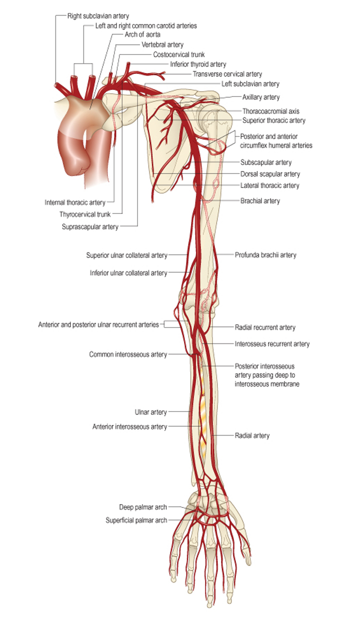
An extensive vascular system supplies blood. Vessels are involved in the transport of oxygen, the nutrition of the tissues of the joint, and are involved in the removal of decay products along with the blood. The shoulder joint is located next to two large arteries, which makes damage dangerous. With a strong displacement of the head or with a fracture of the splinter type, there is a possibility of rupture or compression of the vessels.
If trauma to the shoulder joint has contributed to arm numbness or a strong feeling of weakness, then you should immediately visit a doctor. Such signs indicate a violation of the circulatory process, requiring special medical care.
Auxiliary articular elements of the shoulder
The shoulder joint also has other components in its composition, the condition of which determines the health of the entire shoulder.
- The synovial membrane is a thin layer of tissue that covers the inside of the articular surfaces (except cartilage). This component of the shoulder joint nourishes the bone elements due to the rich vascular network. Also, the synovial layer secretes a special secret that reduces friction in the joint during movement and protects it from premature wear. In some cases, inflammation of the synovial membrane, called synovitis, can occur.
- Periarticular bags are structures responsible for softening the movements of all components of the shoulder and protecting them from wear. Bags are made in the form of pockets with liquid. The inflammation of these bags is called bursitis.
Shoulder research methods
Movements in the shoulder joint are closely related to the mobility of the shoulder girdle. That is why their research is most often carried out at the same time. In addition to X-ray examination, a number of other diagnostic methods are used.
- Physical methods (examination, palpation, tests for the study of active and passive movement in the joint, functional tests).
- Arthroscopy is an invasive method for endoscopic imaging of the components of a joint.
- Thermography - a method based on the analysis of infrared radiation of the body, is used to identify areas of inflammation.
- Ultrasonography is a method of ultrasonic diagnostics of the shoulder joint.
- Radionuclide analysis is a method of studying the human body, based on the introduction of radionuclide particles into the body and the study of their movement and placement in tissues and organs.
- Puncture of the synovial bag - used to study the synovial fluid and identify signs of inflammation.
- Biopsy - used to microscopically study a tissue sample from the articular joint and detect pathology at the cellular level.